The male reproductive system comprises the testes, external genitalia (penis), and accessory ducts such as the rete testis, vasa efferentia, epididymis, and vas deferens. Unlike the female reproductive system, most male reproductive organs are located outside the body. Additionally, the male reproductive system includes accessory glands like the prostate glands, seminal vesicles, and bulbourethral glands.
Male Reproductive System Diagram
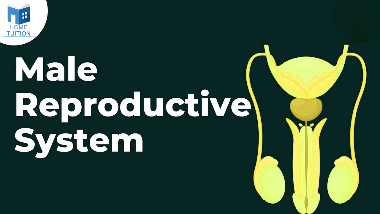
Male Reproductive System
.png)
Male reproductive system comprises of following parts:
Also Read: Human Reproductive System
Testis
They are soft, smooth, pinkish, oval organs. They are housed [present] in a sac like structure called as scrotum. Outer covering is called as as tunicavaginalis. It’s inner covering is called as tunica albuginea. Ingrowths of tunica albuginea are called as septa, that divide the testis into 200-300 lobules.
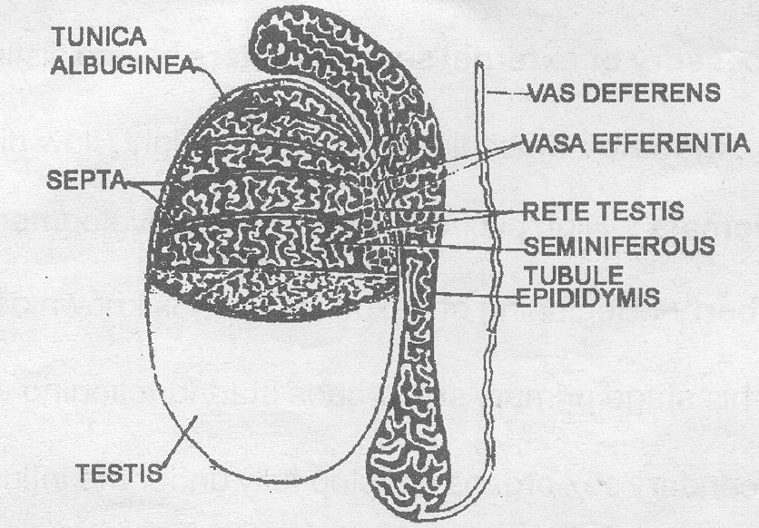
- It also consist of convoluted somniferous tubules.
- These somniferous tubules at one end join to form tubules which open into a network of irregular cavities known as rete testis.
- This rete testis comes out from a dorsal surface of the testis with the help of vesa efferentia.
- This vasa efferentia combines to form a single tube which becomes highly coiled and from epididymis.
- This vasa efferentia combines to form a single tube wihich becomes highly coiled and form epididymis.
- Epididymis peon into a narrow tube vas deferens.
- Somniferous tubules from the spermatogenic tissue of the testis.
- It consists of a germinal epithelial layer at the periphery. Spermatogenesis occurs at the center.
- It forms spermatogonia which grows and form spermatocytes which further grow to form primary spematrocytes, which undergo meiosis to form secondary spermatocytes and then spematids.
- The later (i.e. spermatids) metamorphose into spermatozoa.
- This process of formation of spermatozoa from spermatogonia is called as spermatogenesis.
- These spermatozoa are nourished during the development by nurse cells.
Also Read: Neurons
Scrotum
It is a pouch of pigmented skin arising from the lower abdominal wall and hanging between the legs.
- It is divided internally into two compartments by a muscular partition called as septum scroti.
- Scrotum possesses smooth involuntary dortus muscles.
- Scrotum sac is connected to the abdominal cavity through inguinal canal.
- Function of dortus muscle is to change the position of testis to keep them at proper temperature.
- Scrotum has temperature 1 - 3 lower than body temperature which favours the formation of sperms
Also Read: Pollination
Testes
In humans, a pair of testes is present. The testes are located outside the body in a pouch called the scrotum. They are oval-shaped organs, approximately 4 to 5 cm long and 2 to 3 cm wide. Generally, the left testis hangs slightly lower than the right one.
The primary functions of the testes are:
- Producing testosterone, a male sex hormone.
- Producing sperms through spermatogenesis, which carry a man's genes.
Each testis consists of about 250 testicular lobules or compartments.
Sperms are produced in the seminiferous tubules. Each testicular lobule contains one to three seminiferous tubules, which are lined by two types of cells:
- Spermatogonia or male germ cells, which undergo spermatogenesis to produce sperm.
- Sertoli cells, which provide nutrition to germ cells.
Leydig cells or interstitial cells are present outside the seminiferous tubules in the interstitial spaces. They secrete male sex hormones or androgens, such as testosterone.
Also Read: Fibrous Joints
Male Sex Accessory Ducts
The male sex accessory ducts include the rete testis, vasa efferentia, epididymis, and vas deferens. The seminiferous tubules open into the rete testis, which then leads to the vasa efferentia. From there, the vasa efferentia open into the epididymis, and the epididymis leads to the vas deferens. The vas deferens joins with a duct from the seminal vesicle to form the ejaculatory duct. These ducts are where sperm mature, are stored, and are transported.
Urethra
The urethra is a tube-like structure that connects the urinary bladder to the urinary meatus. In males, it travels through the penis and serves two main functions:
- It is part of the urinary tract, conveying urine from the bladder where semen is ejaculated.
- The prostate, located beneath the bladder, surrounds the urethra. It enlarges with age, and if it grows excessively, it can obstruct urine flow through the urethra, leading to urinary symptoms.
Male Accessory Glands
The male accessory glands consist of the seminal vesicles, prostate, and bulbourethral glands. There is a pair of seminal vesicles present. These vesicles are positioned above the prostate and are connected to the vas deferens, forming the ejaculatory ducts that pass through the prostate. These glands produce a fluid called seminal plasma, which nourishes the sperm. Seminal plasma is rich in fructose, calcium, and various enzymes. It comprises the majority of the semen volume, which is ejaculated during ejaculation.
Frequently Asked Questions
Ans. Produces, maintains, and transports sperm (the male reproductive cells) and protective fluid (semen) Discharges sperm during sex. Produces and secretes male sex hormones are the function of the male reproductive system.
Ans. The 7 male reproductive system includes the penis, scrotum, testes, epididymis, vas deferens, prostate, and seminal vesicles.
Ans. Sperm develop in the testicles.
Ans. The testes serve as the primary sex organs in males. Additionally, the accessory sex organs encompass the prostate gland, seminal vesicles, urethra, and penis.