Share
Report
Question
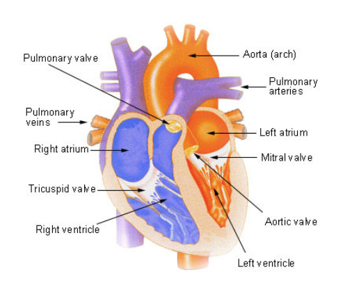
The internal structure of the human adult heart:
It consists of three layers:
- Epicardium: it is made up of fibrous tissue and maintains its shape.
- Myocardium: it is the thickest layer made up of cardiac muscle fiber.
- Endocardium: it is the epithelial lining.
It is divided into two atria, and two ventricles making a four-chambered heart; it prevents the mixing of deoxygenated and oxygenated blood in the body and helps proper pumping of blood.
- Atrium: The upper chambers are termed atria. The left atrium is the upper left side that receives oxygenated blood from pulmonary veins The right atria is the upper right chamber that receives blood from superior and inferior vena cava.
- Ventricles: The inferior chambers are termed ventricles. The left ventricle receives oxygenated blood from the left atrium and releases blood to the aorta. The right ventricle receives blood from the right atria and transfers to the pulmonary trunk.
Pulmonary artery carries deoxygenated blood from the heart to the lungs for oxygenation while the pulmonary vein brings oxygenated blood from lungs to heart.
Aorta is the main artery that carries blood to each and every part of the body.
Vena cava are of two types, superior vena cava which brings blood from the head and neck portion of the body while the inferior vena cava is the main vein that brings blood from lower portion of the body to the heart.
Different septums separate these four chambers of the heart.
Atrioventricular septum: the thick septum which separates atria and ventricle. It consists of a guarded opening by an atrioventricular valve (AV valve).
The AV valve between the right atria and ventricle is a tricuspid valve, made up of three cusps.
The AV valve between the left atria and ventricle is a bicuspid valve. Also called the mitral valve.
Interatrial septum: it is a thin septum that separates the right and left atria.
Interventricular septum: it is a thick septum that the two ventricles and are also attached to the ventricle wall.
Eustachian valve: the EV is present in the upper portion of the inferior vena cava and enters into the right atrium.
Thebesian valve: It is the valve of coronary sinus that is the collection of veins which carries deoxygenated blood to the right atrium.
The valves prevent the backflow of blood. The backflow of blood into the ventricles is prevented by the semilunar valve.
Chordae tendineae: it is the fibrous tissue that maintains the position of AV valves and prevents their opening into the atria.
The connection between the interatrial septum is closed after birth to form fossa ovalis.

Final Answer: The internal structure of the heart is marked by three layers and four chambers separated by septa.
solved
5
wordpress
4 mins ago
5 Answer
70 views
+22

Leave a reply